
What a 2D X-ray does not tell you
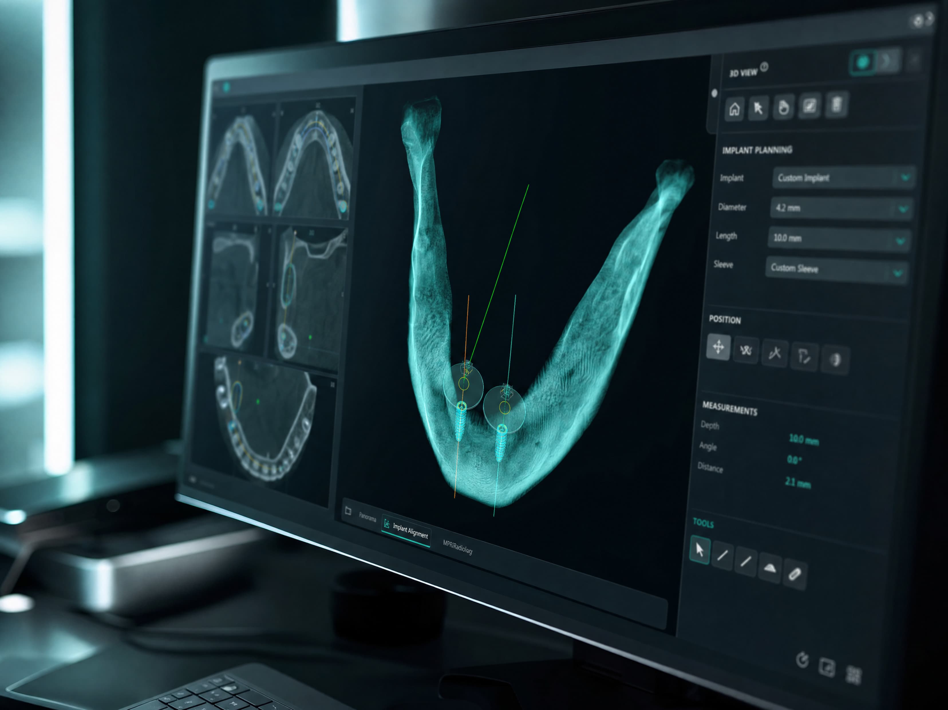
Digital implant planning takes a CBCT scan of the jaw, merges it with an intraoral or model scan of the teeth, and lets the implant be positioned virtually against the real bone, nerve, and sinus anatomy before surgery starts, then produces a surgical guide that carries that exact plan into the mouth. The result is a plan built backward from the final crown, not just a spot where a scan shows enough bone.
A periapical or panoramic X-ray gives you a flat, two-dimensional picture of a three-dimensional problem. You can see roughly where bone is, but not how thick it is buccal to lingual, not exactly where the inferior alveolar nerve canal sits relative to a proposed implant site, and not the true floor of the maxillary sinus. Placing an implant based on that alone means working with an incomplete picture and leaning on experience to fill the gaps.
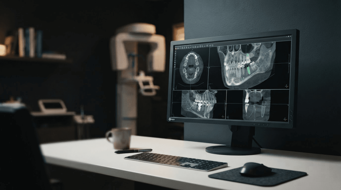
CBCT (cone beam computed tomography) closes that gap. It gives you an actual 3D volume of the jaw: bone height, width, and density at the exact site, the nerve canal's real path, sinus anatomy, and the position of adjacent roots. None of that is guesswork anymore. It is measured.
What makes a CBCT scan actually usable for planning
Not every CBCT scan is good enough to plan from, and the scan itself is where a lot of avoidable problems start. A few things determine whether a scan is actually usable:
Field of view (FOV) matched to the case. A small FOV centered tightly on one or two teeth is fine for a single implant, but it will miss the sinus, the opposing arch, or the full nerve path on a case that needs that context. A limited FOV taken to reduce radiation dose can end up costing more once a second, wider scan has to be taken to see what the first one cut off.
Voxel size appropriate to the anatomy being measured. A voxel size that's too large blurs exactly the detail that matters most: the cortical border of the nerve canal, fine trabecular bone changes, and the true thickness of a thin buccal plate. For implant planning specifically, a smaller voxel size in the region of interest is worth the modest increase in scan time.
Minimal motion artifact. Patient movement during the scan shows up as streaking or doubling in the image, and it degrades measurement accuracy exactly where precision matters most. A short scan time and clear instructions to the patient beforehand reduce this more than any software correction after the fact.
Metal artifact from existing restorations. Crowns, posts, and old amalgam create scatter that can obscure adjacent anatomy on the same scan. This does not make a scan unusable, but a planner needs to know it is there and plan around the affected region rather than trust the artifact-corrupted area at face value.
A scan that gets these basics right is what makes everything downstream, from the virtual implant placement to the guide design, actually trustworthy rather than a precise-looking plan built on an imprecise foundation.
What the digital planning workflow actually involves
A CBCT scan on its own is useful, but the real value shows up when it is combined with a proper planning workflow:
- The CBCT scan (DICOM data) and an intraoral or model scan (STL) are merged into one 3D model, so you are working with both accurate bone anatomy and the exact shape of the teeth and soft tissue.
- The implant is placed virtually in planning software, positioned against the real bone volume and the nerve canal, sinus floor, and adjacent structures.
- Critically, the implant position is checked against where the final crown needs to sit, not just against where there happens to be bone. This is what "prosthetically-driven" planning means, and it is the difference between an implant that is surgically placed and one that is also restoratively sound.
- Once the position is confirmed, a surgical guide is designed and produced, translating the plan into a physical tool that controls angulation and depth during surgery.
Why prosthetically-driven planning matters more than it sounds
It is possible to place an implant in a spot with plenty of bone that still turns into a restorative problem: an angulation that forces a screw access hole through the middle of the facial surface, or a position that makes a hygienic, cleansable crown contour difficult to achieve. Planning that starts from the CBCT bone data but also references the intended final restoration catches this before surgery, not after the crown is already being fabricated around a compromised implant position.
The specific surgical risks this workflow reduces
This is not a theoretical benefit. Digital, CBCT-based planning directly reduces a specific, known set of surgical risks:
Nerve injury. The inferior alveolar nerve canal is visible and measurable in 3D, so the planned implant depth can maintain a real, deliberate safety margin instead of an estimated one.
Sinus perforation. Maxillary sinus floor position and any pneumatization are visible before you ever pick up a handpiece, so posterior maxillary implants can be planned around the actual anatomy rather than average anatomy.
Inadequate primary stability. Bone density at the exact site is visible, which affects both implant selection and whether a site needs grafting first, decided before surgery rather than discovered during it.
Misangulation. A surgical guide controls the drilling angle and depth mechanically, which removes a meaningful amount of the variability that comes from placing freehand off a 2D image and clinical judgment alone.
Surgical guides and what they change in the operating chair
A guide produced from the digital plan controls both angulation and depth during osteotomy prep, which is where freehand placement has the most room to drift from the plan. This does not remove the surgeon's judgment from the procedure. It gives that judgment a physical constraint that matches the plan made with full 3D information, instead of leaving the final angulation to be corrected by eye in real time. For multi-implant or full-arch cases, this consistency across several sites is where the guide's value is most obvious, since small angulation differences between implants compound into much bigger prosthetic problems.
Guide design considerations that affect accuracy
Not all surgical guides are built the same way, and the support type matters as much as the plan behind it:
Tooth-supported guides rest on remaining teeth and are generally the most stable and accurate option when enough teeth are present to seat the guide reliably in one position.
Mucosa-supported guides, used in edentulous or partially edentulous spans, rely on soft tissue for support and are more sensitive to soft tissue compressibility and how precisely the guide seats. These usually benefit from a fixation pin to stop the guide from lifting or rocking during drilling.
Bone-supported guides, typically used after flap reflection, offer a very stable reference but require exposing the bone, which changes the case from flapless to open surgery.
Sleeve height and drill key system also affect real-world accuracy. A taller sleeve gives the drill more guidance but needs enough vertical space to fit, which is not always available in a limited interocclusal space. The guide should always be tried for a passive, fully-seated fit before it goes anywhere near a handpiece. A guide that rocks even slightly transfers that same error directly into the osteotomy, which defeats the purpose of planning it precisely in the first place.
A common misconception worth addressing directly
Some clinicians hesitate to add a CBCT and digital planning step because it feels like it slows the workflow down, or because they assume the radiation dose is a significant added burden on the patient. In practice, a single CBCT scan's effective radiation dose is generally in the same range as a full-mouth series of traditional radiographs, and considerably lower than a medical CT scan, while the diagnostic information it provides for implant planning specifically is far more complete. The time added to planning is also typically recovered, and then some, in reduced chair time and fewer surprises during surgery.
Where cases commonly go wrong
Most problems in digital implant planning trace back to a small number of avoidable mistakes, not to the technology itself:
Planning from CBCT alone, without merging an intraoral scan. CBCT soft-tissue and tooth-surface detail is far less accurate than a proper optical scan. Skipping the merge step and estimating tooth position from the CBCT alone reintroduces exactly the kind of imprecision digital planning is supposed to remove.
Planning surgically first and checking the restoration second. A position chosen purely because there is bone there, without checking it against where the final crown needs to emerge, is the single most common source of a technically successful but restoratively difficult case.
Never verifying the guide's fit before surgery day. A guide that was designed correctly can still be manufactured with a slight inaccuracy, or a model can shift between design and printing. Trying the guide in the mouth, or on a printed model, before the day of surgery catches this while there is still time to reprint it.
Treating the plan as final regardless of what surgery reveals. A digital plan is built on the best information available before the flap is raised. If bone quality or an anatomical detail looks different once surgery is underway, the plan is a strong starting point, not a script that overrides clinical judgment in the room.
What to have ready if you are sending a case out for planning
If implant planning is being handled by an outside provider rather than entirely in-house, the case moves faster when you send it complete the first time: the CBCT DICOM export, an intraoral or model scan (STL), the planned restoration type and number of units, any specific prosthetic requirements from the restoring dentist, and relevant medical history that could affect bone quality or healing. A planning service that is used to working from these files should be able to turn around a proposed implant position, guide design, and a written rationale you can review before anything is manufactured.
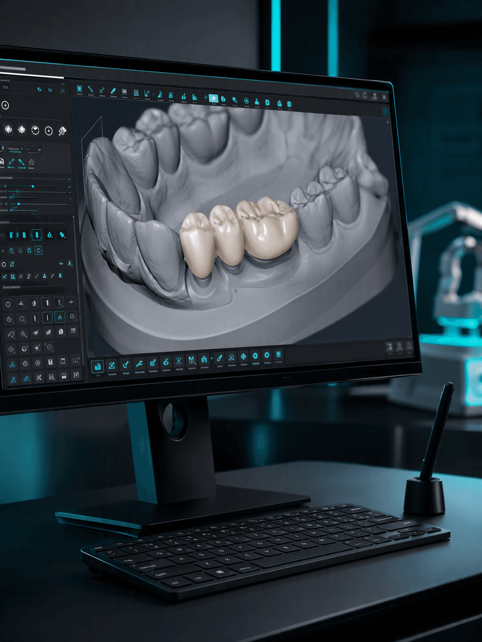
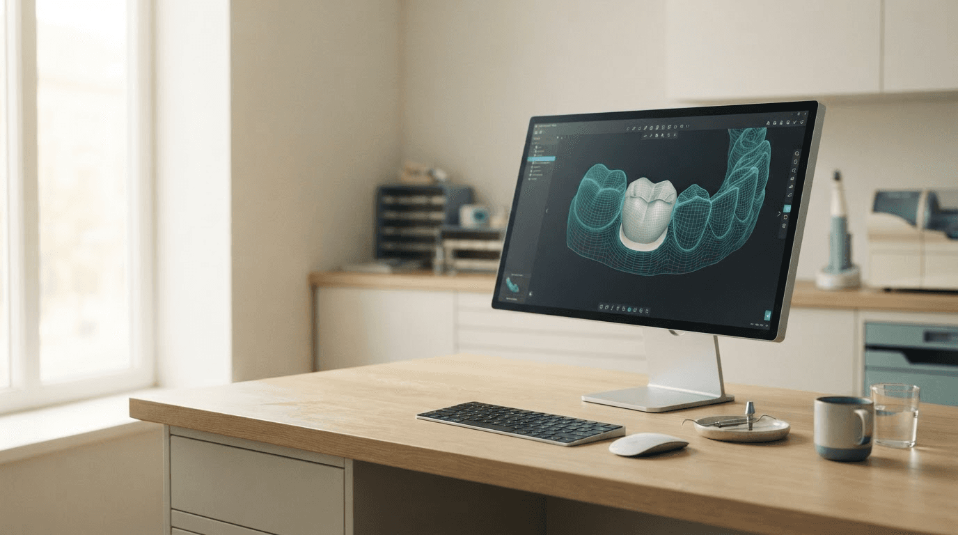
If the final restoration itself still needs to be designed, that is a separate but related step. An Exocad design service can produce the crown or bridge file from the same scan data once the implant position is confirmed.
The point of all this
None of this replaces clinical judgment. A surgeon still decides whether a case is appropriate for implants at all, still manages the soft tissue, still handles whatever comes up mid-procedure that no scan could have predicted. What CBCT-based digital planning does is remove the parts of the decision that do not need to be guesswork. Bone volume, nerve position, sinus anatomy, and implant angulation are all things that can be measured and planned precisely before surgery starts. Using that information is not a shortcut. It is simply operating with the full picture instead of a partial one.
If you want a second set of eyes on a case before committing to surgery, Denta Dynamics' implant planning service is priced at $15 per tooth being planned, with a full plan and surgical guide design, if you need one, turned around within 24 hours.